
Capsule Endoscopy
Capsule endoscopy is a non-invasive diagnostic procedure designed to visualize the small intestine, a segment of the gastrointestinal tract that is difficult to assess using conventional endoscopy or colonoscopy. The small bowel plays a crucial role in digestion and nutrient absorption, and diseases affecting this region can be challenging to diagnose due to its length and location. Capsule endoscopy has transformed the evaluation of small bowel disorders by providing detailed, continuous imaging without the need for sedation or invasive instrumentation.
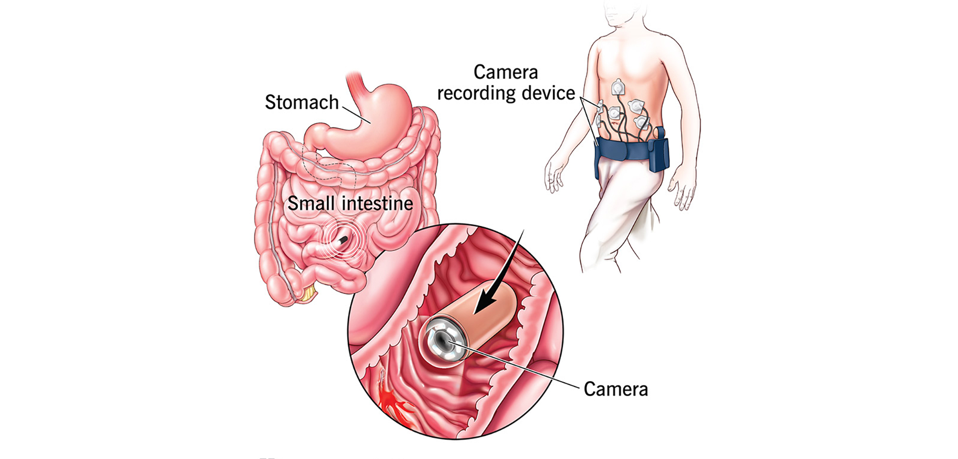
The procedure involves swallowing a capsule approximately the size of a large vitamin pill. This capsule contains a miniature camera, light source, battery, and wireless transmitter. As the capsule travels naturally through the digestive tract, it captures thousands of high-resolution images of the small intestine, which are transmitted to a data recorder worn by the patient. These images are later reviewed by a gastroenterologist to identify abnormalities.
Capsule endoscopy is primarily indicated for evaluation of unexplained gastrointestinal bleeding when standard endoscopy and colonoscopy fail to identify a source. It is also widely used in suspected Crohn’s disease, particularly when disease is confined to the small intestine. Other indications include suspected small bowel tumors, polyps, celiac disease, chronic abdominal pain, and iron-deficiency anemia of unknown origin.
One of the major advantages of capsule endoscopy is its patient-friendly nature. The procedure does not require sedation, intubation, or hospital admission. Patients can continue most of their daily activities while the capsule records images. Preparation is relatively simple and typically involves fasting and, in some cases, bowel cleansing to improve visualization.
Capsule endoscopy provides a comprehensive view of the small bowel mucosa, enabling detection of ulcers, inflammation, bleeding lesions, strictures, and tumors. It is particularly valuable in identifying subtle lesions that may be missed on radiological imaging. While the capsule does not allow tissue biopsy or therapeutic intervention, the diagnostic information it provides guides further targeted investigations or treatment.
After the capsule completes its journey and is naturally passed in the stool, the recorded data is analyzed. Results help determine whether additional procedures, such as balloon-assisted enteroscopy, medical therapy, or surgery, are required. Capsule retention is rare but carefully assessed through patient selection and pre-procedure imaging when necessary.
Capsule endoscopy has become an essential diagnostic tool in modern gastroenterology. Its ability to safely and effectively evaluate the small intestine improves diagnostic accuracy, reduces delays in treatment, and enhances overall management of complex gastrointestinal disorders.
Quick Contact
If you have any questions simply use the following contact details.

Working Hours
-
Out-patient Department
Monday to Saturday 08:00 AM - 09:00 PM
Sunday 10:00 AM - 06:00 PM
-
Emergency Department & Pharmacy
Sunday to Saturday 24x7


Book Appointments, Inquire, or Manage Your Care Easily – Get in Touch via
 04 406 3000
  04 406 3000
Quick Links
Legal
Working Hours
-
Out-patient Department
Monday to Saturday | 8 AM to 9 PM
Sunday | 10 AM to 6 PM
Emergency Department & Pharmacy | 24x7
Connect with us!


©2026 IMH. All Rights Reserved. Ministry of Health License No : TEBMJIU0-060622